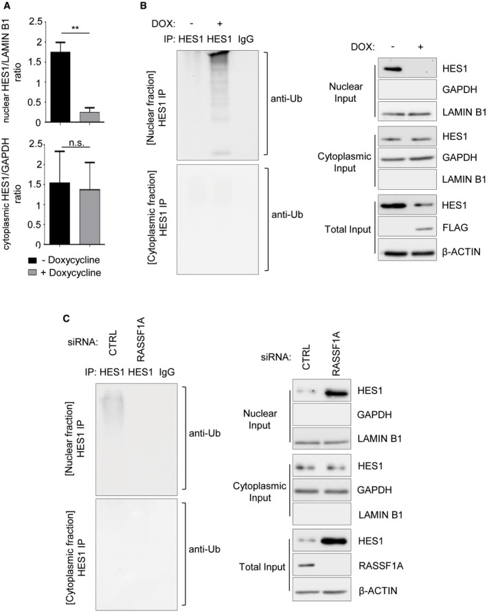
Figure EV2. RASSF1A‐mediated ubiquitination of HES1.

- Densitometry on Fig 1G showing that only nuclear HES1 levels are significantly reduced upon RASSF1A induction, based on the nuclear HES1/LAMIN B1 ratio. Cytoplasmic HES1 levels remain unaffected based on the cytoplasmic HES1/GAPDH ratio.
- In vivo ubiquitination assay in HES1 immunoprecipitates from nuclear and cytoplasmic fractions of U2OS cells Tet‐On inducibly expressing FLAG‐RASSF1A versus Control. Doxycycline (DOX) was used at a concentration of 0.5 μg/ml for 24 h. Immunoprecipitates and Input lysates are probed with displayed antibodies.
- In vivo ubiquitination assay in HES1 immunoprecipitates from nuclear and cytoplasmic fractions of U2OS cells transfected with either siCTRL or siRASSF1A. Immunoprecipitates and Input lysates are probed with displayed antibodies.
Data information: **P < 0.01, of Student’s t‐test. Error bars indicate s.e.m. Data shown are representative of three biological replicates (n = 3).
Source data are available online for this figure.
