Figure 3.

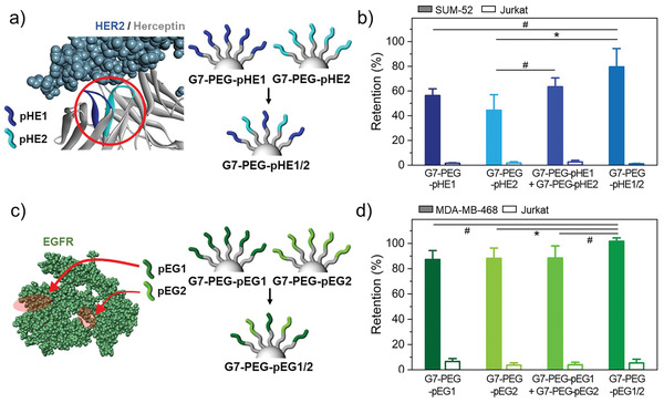
Dendrimer‐PEG0.5k surfaces coimmobilized with multiple peptide sequences that bind to different sites within a single protein: a) Schematic illustration of G7‐PEG surfaces immobilized with two different peptide sequences that bind to different sites of HER2 protein, pHE1 and pHE2. b) Retention of surface‐bound SUM‐52 and Jurkat cells on HMA surfaces consisting of G7‐PEG0.5k‐pHE1, G7‐PEG0.5k‐pHE2, a mixture of the two DPCs, or G7‐PEG0.5k‐pHE1/2 upon washing. c) Schematic illustration of G7‐PEG0.5k surfaces immobilized with two different peptide sequences that bind to different sites of EGFR protein, pEG1 and pEG2. d) Retention of surface‐bound MDA‐MB‐468 and Jurkat cells on HMA surfaces consisting of G7‐PEG0.5k‐pEG1, G7‐PEG0.5k‐pEG2, a mixture of the two DPCs, or G7‐PEG0.5k‐pEG1/2 upon washing. All washing steps for cell retention assays were performed at a flow rate of 50 µL min−1, which corresponds to a shear stress of 0.36 dyne cm−2. Significance levels are indicated as # p < 0.10, * p < 0.05, ** p < 0.01, and *** p < 0.001, which are analyzed using Student's t‐test.
