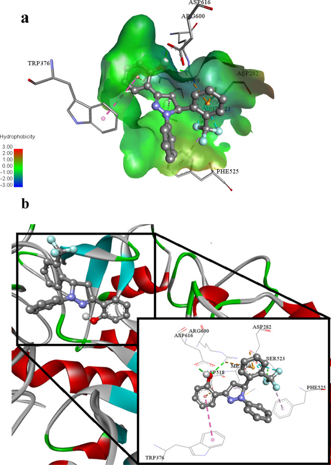
Figure 14.
(a) Putative binding interactions of ligand 2o against α-glucosidase. (b) Interactions of the ligand 2o in 3D space. Interactions with specific amino acid residues are shown in the box. The 3D ribbon represents the enzyme-stick model of the lowest energy conformers of the inhibitor 2o along with amino acids of α-glucosidase interacting with it.

