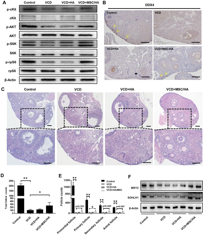
Fig. 2.
MSC/HA transplantation activated the PI3K-AKT pathway and increased follicle numbers in POI mouse model. a Western blot showing the activation of the PI3K-AKT pathway in the control, VCD, VCD + HA, and VCD + MSC/HA groups 4 days after transplantation. b Immunostaining showing the expression of DDX4 in ovaries 1 week after transplantation in the four groups. The yellow arrows indicate primordial follicles. Scale bars = 50 μm. c H&E staining of ovaries from the four groups at 10 weeks after transplantation. Scale bars = 100 μm. d Numbers of total follicles and e follicles at various stages were counted at 10 weeks after transplantation and compared with VCD group (n = 8 for each group). *P < 0.05, **P < 0.01. f Western blot showing the expression of germ cell markers MSY2 and SOHLH1 in ovaries from the four groups at 10 weeks after transplantation

