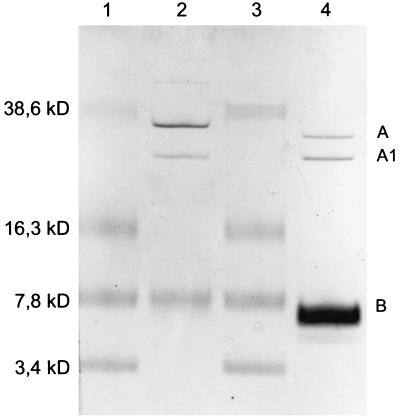
FIG. 1.
SDS-PAGE demonstrating the purity of the specific batch of toxins used in the present study. Shown are the A subunit with the A1 fragment and the B subunit of Stx2 (lane 2) and Stx1 (lane 4) visualized after silver staining according to the method of Merril et al. (29). Lanes 1 and 3, low-molecular-weight protein standard (Rainbow unstained; Bio-Rad).

