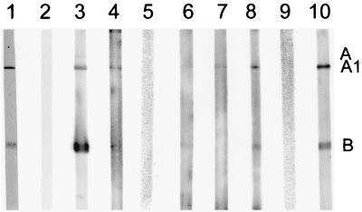
FIG. 2.
Western immunoblot of human and rabbit sera against Stx1 as antigen (lanes 2 to 10). Lane 1 shows a strip of the PVDF membrane after blotting of Stx1, showing the A1 fragment and the B subunit visualized after Coomassie blue staining. Immunoblot strips from five human sera collected from HUS patients were used as standard control sera for the Stx1 WBA, two negative (lanes 2 and 5, nonreactive) and three positive (lane 3, reactive to the A subunit [weak], the A1 fragment, and the B subunit; lane 4, reactive to the A1 fragment; lane 6, reactive against both the A1 fragment and the B subunit [very weak]). Positive human sera from HUS patients (lane 7, reactive against the Stx1 A1 fragment [weak]; lane 8, reactive against the A1 fragment and the B subunit), preimmune rabbit serum (lane 9, nonreactive), and Stx1 immune rabbit serum at a dilution of 1:5,000 (lane 10, reactive against the Stx1 A1 fragment and the B subunit) are shown.

