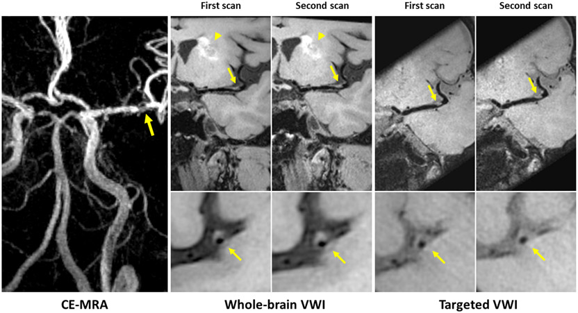
Figure 3.
Representative 3D scan and rescan images of a patient with a left basal ganglia infarct (arrowhead). Contrast-enhanced magnetic resonance angiography (CE-MRA) shows severe stenosis of left middle cerebral artery. Both 3D whole-brain and targeted vessel wall imaging (VWI) depict an eccentric atherosclerotic plaque at the corresponding location (arrows) with good delineation of the plaque on two scans. However, the outer boundary is difficult to assess on the targeted image due to insufficient cerebral spinal fluid (CSF) suppression.

