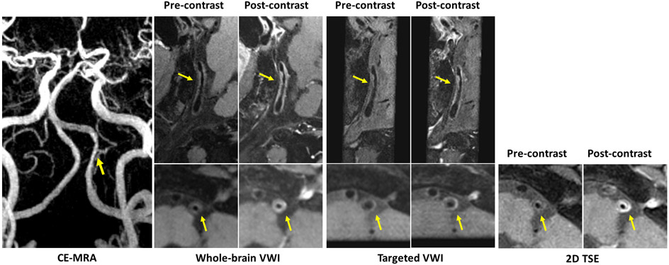
Figure 4.
Representative 3D and 2D images with (post-contrast) and without contrast agent (pre-contrast) for a patient with a severe stenosis of left vertebral artery on contrast-enhanced magnetic resonance angiography (CE-MRA). A long atherosclerotic plaque detected at the corresponding location (arrows) on both 3D whole-brain and targeted vessel wall imaging (VWI) is considered as vulnerable plaque with obvious enhancement on post-contrast images. The post-contrast whole-brain image exhibits similar enhancement of the plaque to the 2D turbo spin echo (TSE) image, but more enhancement than the post-contrast targeted image.

