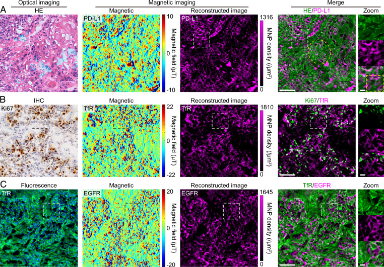
Fig. 6.
Correlated magnetic and optical imaging in lung tumor tissues. (A) Correlated HE staining and IMM in the same tissue section. Hematoxylin and eosin stained the tissue’s cellular structure, simultaneously producing strong fluorescence signals (SI Appendix, Fig. S12), which significantly reduced the contrast of the NVs’ CW spectrum (SI Appendix, Fig. S13). Nevertheless, the robust IMM resisted the impact of fluorescence from hematoxylin and eosin. Although the magnetic image shows a slightly reduced signal-to-noise ratio, it still clearly resolves the distribution of PD-L1. The yellow ellipse and box in the merged images indicate squamous carcinoma cells and immune cells, respectively. (B) Double labeling of Ki67 and TfR by IHC and immunomagnetism in the same section, respectively. In IHC, DAB immunostained Ki67 proteins and hematoxylin stained cell nuclei. Again, the IMM resisted the impact of DAB and hematoxylin, and we obtained a high-quality magnetic image. (C) Double labeling of TfR and EGFR by IF and immunomagnetism in the same section, respectively. Alexa Fluor 488 in the green channel represents TfR, which was labeled via the routine immunofluorescence procedure. Shown are DAPI-stained cell nuclei. Both imaging methods work well without disturbing each other. The HE and DAB signals were recolored with the green color, and then the optical images were merged with the reconstructed IMM images. These results were confirmed in three (IMM and HE), four (IMM and IHC), and two (IMM and IFM) samples in independent experiments. (Scale bars, 100 μm and 20 μm [zoom].)

