Fig. 3.
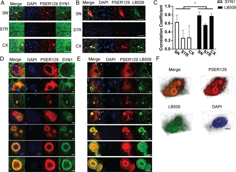
Intracellular distribution of BAR-PSER129–labeled pathology in the synucleinopathy brain. Formalin-fixed floating brain sections from human synucleinopathy cases were fluorescently labeled by substituting fluorescent tyramides for biotinyl tyramine in the standard BAR protocol. (A) Sections were dual labeled for total alpha-synuclein (SYN1) and phosphoserine 129 alpha-synuclein (PSER129). Representative confocal images are from brain regions of interest. (Scale bars, 50 μm.) (B) Sections dual labeled for the LP marker (LB509) and PSER129. Representative confocal images are from brain regions of interest. (Scale bars, 50 μm.) (C) Colocalization analysis of dual-labeled brain sections. Correlation coefficient was calculated for confocal images acquired under each experimental condition. Subcellular distribution of PSER129 and total (D) alpha-synuclein (SYN1) and (E) LB509 in LP-bearing perikarya. Images depict diversity of LP morphology, including granular morphology to compact circular type. (Scale bars, 5 μm.) (F) Three-dimensional projection images of multiple Lewy bodies in the cytoplasm of a cell labeled with PSER129 and LB509. (Scale bars, 10 μm.) Nuclei were stained in all sections using DAPI. Two-way ANOVA *P < 0.05, n = 3.

