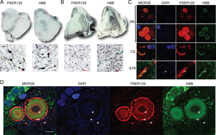
Fig. 6.
BAR-PSER129–identified protein hemoglobin beta (HBB) interaction with LP. Experiments were performed to confirm BAR-PSER129 enrichment of LP. Formalin-fixed human brain sections were immunolabeled for PSER129 and HBB. Nickle DAB detection of immunolabeled PSER129 and HBB in both (A) the human SN and (B) the section containing the STR. (SN scale bars, 20 μm; STR scale bars, 50 μm.) Top of A and B depicts whole section scans obtained using a 4× objective. Bottom is a representative image obtained with a 20× objective. PSER129 and HBB were labeled with fluorescent tyramines and imaged using confocal microscopy. (C) Single z-plane images acquired with a 60× objective showing labeling in the SN, STR, and adjacent cortical regions (CX). SN images depict labeling of a cell containing a single Lewy body, granular-type pathology, and multiple Lewy bodies. (SN scale bars, 5 μm; CX and STR scale bars, 1 μm.) White arrows depict DAPI signal within the PSER129/HBB-labeled Lewy neurite. (D) Single z-plane confocal image showing distribution of HBB and PSER129 in the Lewy body. White arrows highlight DAPI signal in proximity and within the Lewy body (n = 2, case 3 and case 4). Scale bar, 5 μm.

