
Figure 1.
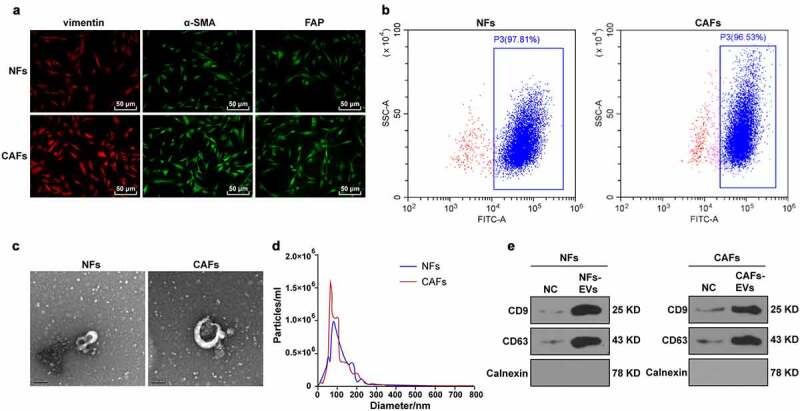
Identification of CAFs and EVs. The CAFs and NFs were successfully isolated from COAD tissues and adjacent normal tissues of patients undergoing colon cancer surgery. (a) Immunofluorescence was used to observe the morphology of CAFs and the expressions of surface markers vimentin, α-SMA, and FAP; (b) The purity of NFs and CAFs was detected using flow cytometry. The EVs were extracted. (c) The morphology of EVs was observed by TEM; (d) The size distribution of EVs was analyzed using NTA; (e) The expressions of CD9, CD63, and Calnexin were analyzed using WB.