
Figure 1. The chytrid fungus Batrachochytrium dendrobatidis is an early branching fungus with an archetypal chytrid life cycle and animal-like and fungal-like actin structures.
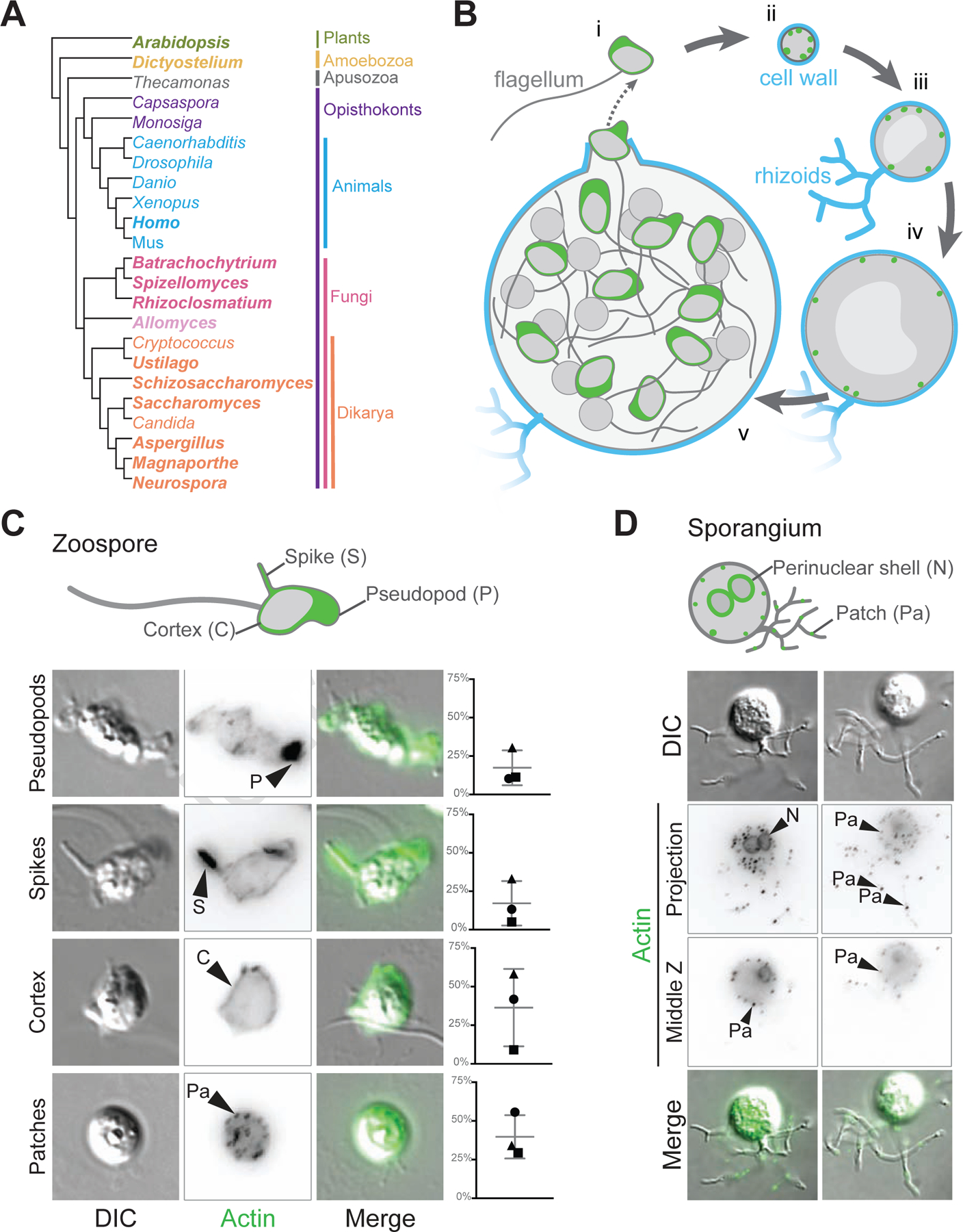
(A) This cladogram shows the relationships between representative genera of major eukaryotic groups. Chytrids are represented by Batrachochytrium, Spizellomyces, Rhizoclosmatium, and Allomyces (magenta, lavender), diverging before the diversification of Dikarya (orange), and are in a sister clade to animals (cyan). Bold type indicates genera used for the majority of the homologous sequence analyses in this paper. (B) In vitro life cycle of Batrachochytrium dendrobatidis (Bd). Bd has a motile stage known as a zoospore (i) with a flagellum made of microtubules, no cell wall, and can crawl using actin (green) based protrusions. Zoospores encyst and build a cell wall (cyan), this stage is referred to as a germling (ii). The germling grows in size, becoming a sporangium. Sporangia develop hyphal-like structures called rhizoids used for nutrient uptake and undergo synchronous rounds of mitosis (iii-iv) before cellularization and release of the next generation of zoos ores (v). This life cycle takes approximately three days in laboratory culture conditions. (C) Representative examples of zoospores (DIC: grey) and the phalloidin stained actin Z-projections of cells at this stage (inverted, black), with an overlay of the two (actin, green). Actin structures in Bd zoospores are: actin-filled pseudopods (P), actin-filled spikes (S), cortical actin (Co), and actin patches (Pa). Graphs on the right indicate the raw percent of cells with the phenotype in 3 independent experiments, shapes here match the shapes for the replicates in Figure 6. (D) Representative examples of Bd sporangia (DIC: grey) and the phalloidin stained actin structures at this stage (inverted, black; both a max intensity z projection and a single slice), with an overlay of the DIC and fluorescence (actin, green). Actin structures in sporangia are: actin patches (Pa), and perinuclear actin shells (N). See also Figure S1.