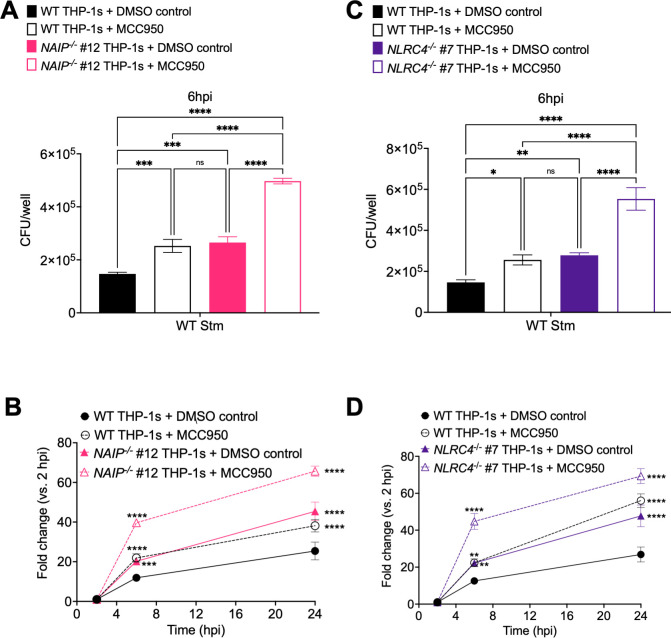
Fig 5. The NAIP/NLRC4 and NLRP3 inflammasomes control Salmonella burdens within human macrophages.
WT, NAIP-/- (A, B), and NLRC4-/-(C, D) THP-1 monocyte-derived macrophages were primed with 100 ng/mL Pam3CSK4 for 16 hours. One hour prior to infection, cells were treated with 1 μM MCC950, a chemical inhibitor of the NLRP3 inflammasome, or DMSO as a control. Cells were then infected with PBS (Mock) or WT S. Typhimurium at an MOI = 20. Cells were lysed at the indicated time points and bacteria were plated to calculate CFU. (A, C) CFU/well of bacteria at 6 hpi (B, D) Fold change in CFU/well of bacteria at indicated time point, relative to 2 hpi CFU/well. ns–not significant, ***p < 0.001, ****p < 0.0001 by Dunnett’s multiple comparisons test (A, C) or Tukey’s multiple comparisons test (B, D). Error bars represent the standard deviation of triplicate wells from one experiment. Data shown are representative of at least three independent experiments.

