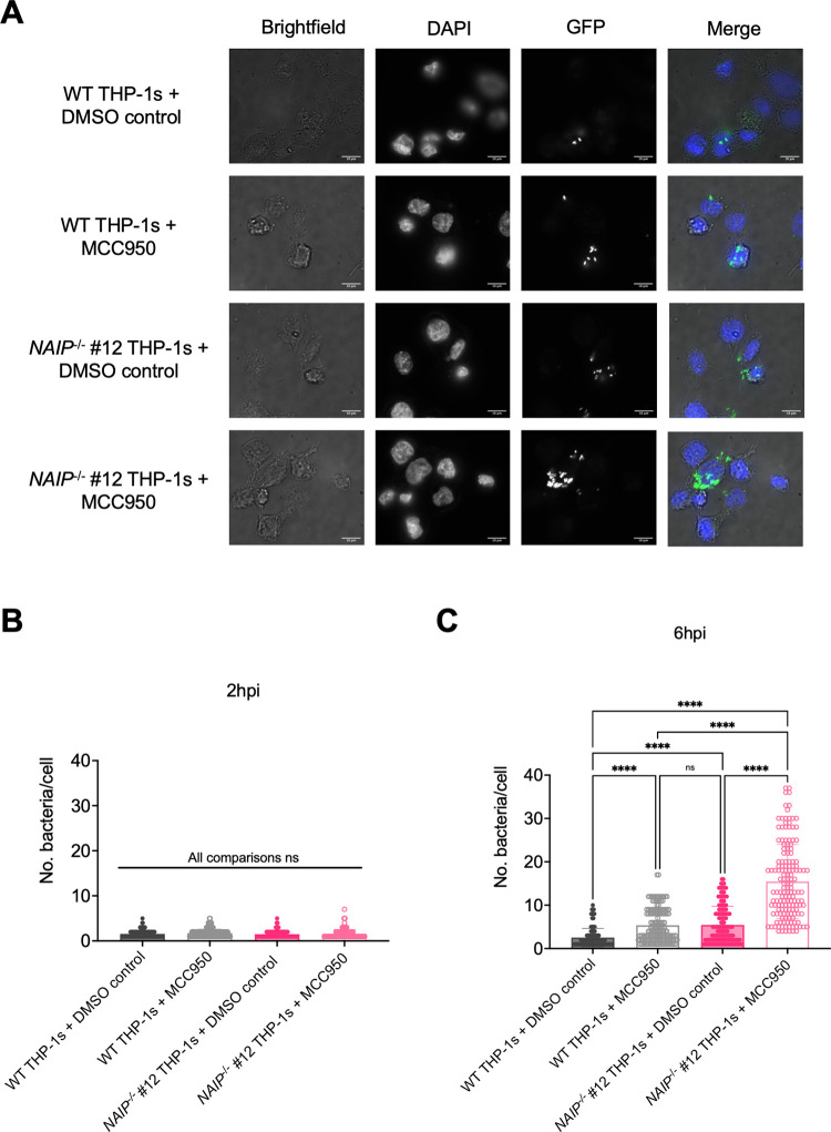
Fig 6. The NAIP/NLRC4 and NLRP3 inflammasomes control Salmonella replication within human macrophages.
WT and NAIP-/- THP-1 monocyte-derived macrophages were seeded on glass coverslips and primed with 100 ng/mL Pam3CSK4 for 16 hours. One hour prior to infection, cells were treated with 1 μM MCC950, a chemical inhibitor of the NLRP3 inflammasome, or DMSO as a control. Cells were then infected with PBS (Mock) or WT S. Typhimurium expressing GFP at an MOI = 20. Cells were fixed at the indicated time points and stained for DAPI to label DNA (blue). The proportion of infected cells containing GFP-expressing Stm (green) and the number of bacteria per cell were scored by fluorescence microscopy. (A) Representative images from 6hpi are shown. Scale bar represents 10 μm. (B, C) Each small dot represents one infected cell. 150 infected cells were scored for each condition (50 infected cells per coverslip). Bars represent the mean from each condition. (B) Number of bacteria/cell at 2 hpi. (C) Number of bacteria/cell at 6 hpi. ns–not significant, ****p < 0.0001 by Tukey’s multiple comparisons test (B). Data shown are representative of at least three independent experiments.

