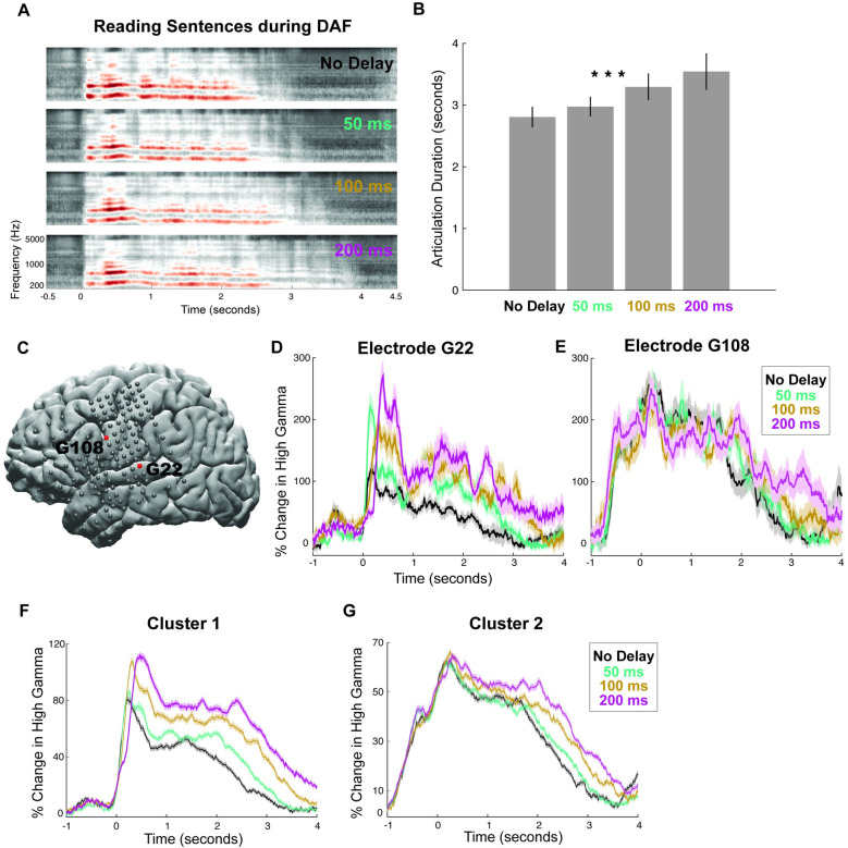
Fig 3. Behavioral and neural responses during sentence reading with DAF.
(A) Speech spectrogram of a single participant articulating sentences during DAF conditions showing a marked increase in articulation duration. (B) Mean articulation duration of sentences during DAF conditions averaged across participants showing a significant effect of duration. Error bars show SEM over participants. (C) Cortical surface model of the left hemisphere brain of a single participant. Gray circles indicate the implanted electrodes. Red highlighted electrodes are located on the STG (G22) and on the vPreCG (G108). (D) High gamma responses in an auditory electrode (G22) to articulation of sentences during DAF conditions (color coded). Shaded regions indicate SEM over trials. (E) High gamma responses in a motor electrode (G108) to articulation of sentences during DAF conditions (color coded). Shaded regions indicate SEM over trials. (F) High gamma responses to articulation of sentences during DAF conditions averaged across electrodes in Cluster 1. Shaded regions indicate SEM over trials. (G) High gamma responses to articulation of sentences during DAF conditions averaged across electrodes in Cluster 2. Shaded regions indicate SEM over trials. The underlying data can be found in https://github.com/flinkerlab/DelayedAuditoryFeedback. DAF, delayed auditory feedback; SEM, standard error of the mean; STG, superior temporal gyrus; vPreCG, ventral precentral gyrus.

