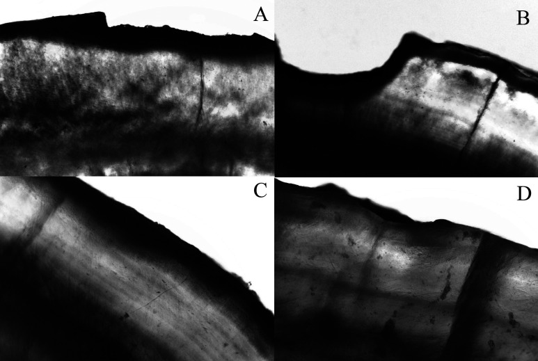
Figure 1.
Artificial enamel caries formation adjacent to bonding area. Specimens were immersed in water and photographed using polarized light microscopy under 10× magnification. The photomicrography shows representatives images of each group. (A,B) Control group showing enamel loss pattern. (C) Dentifrice/dentifrice and mouth rinse pattern of demineralization with no visual mineral loss. (D) Mouth rinse pattern of demineralization presenting low visual mineral loss.

