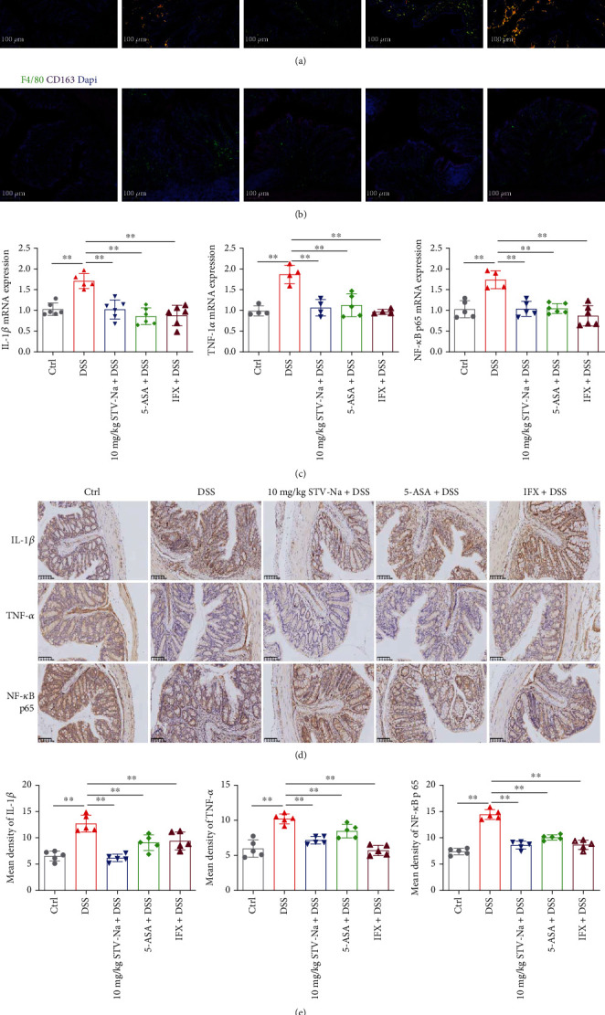
Figure 8.

STV-Na regulated macrophage polarization by inhibiting the secretion of proinflammatory mediators and NF-κB/p65 pathway activation. (a, b) Immunofluorescence staining was performed using anti-CD163, anti-CD86, and anti-F4/80 antibodies to stain colonic F4/80+CD86−CD163− macrophages (M0), F4/80+CD86+CD163− macrophages (M1), and F4/80+CD86−CD163+ macrophages (M2). Nuclear visualization was enabled with Dapi staining. Images show F4/80 (green), CD86 (red), CD163 (orange-red), and Dapi (blue). Scale bar, 100 μm. (c) qRT-PCR was used to evaluate IL-1β (c1), TNF-α (c2), and NF-κB/p65 (c3) mRNA expressions. (d) Immunohistochemical (IHC) analysis was used to evaluate IL-1β, TNF-α, and NF-κB/p65 protein expressions in colonic tissues. Scale bar, 100 μm. (e) The mean density values of IL-1β (e1), TNF-α (e2), and NF-κB/p65 (e3) were measured. Data is depicted in terms of mean ± SD. n = 5 mice per group. An unpaired two-tailed Student's t-test or one-way ANOVA, followed by Tukey's post hoc analysis, was used to analyze the data. ∗P < 0.05 and ∗∗P < 0.01 versus the DSS group.
