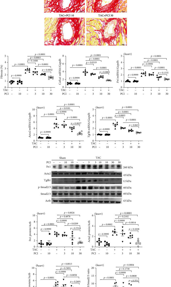
Figure 4.

PCI34051 alleviates cardiac fibrosis in transverse aortic constriction (TAC) mice through the TGF-β1-Smad2/3 pathway. (a, b) Picrosirius red staining of mouse cardiac tissues; representative images and quantification are shown. Scale bar = 50 μm. The expression levels of the fibrosis marker genes Col1a1 (c), Fn1 (d), Acta2 (e), and Tgfb1 (f) were determined using quantitative real-time polymerase chain reaction (n = 5–6 per group). (g) The cardiac expression levels of Fn1, Acta2, Tgfb1, p-Smad2/3, and Smad2/3 were analyzed using western blotting. Actb was used as a loading control. Representative blots are shown. (h–k) Quantification of Fn1, Acta2, Tgfb1, and p-Smad2/3 to Smad2/3 (n = 5–6 per group).
