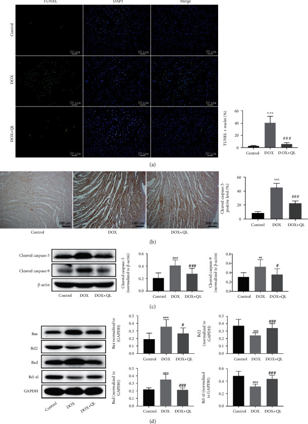
Figure 5.

QL inhibited DOX-induced cardiomyocyte apoptosis in rats under DOX exposure for 28 days. (a) TUNEL staining of cardiomyocytes in three groups. Positive nuclei were stained in green, and nuclei were stained in blue (n = 7 per group, 300 dpi, scale bar: 50μm). (b) Representative immunohistochemical images of cleaved caspase-3 expression in heart tissues in three groups (n = 4 per group, 300 dpi, scale bar: 100μm). (c, d) The expression levels of cleaved caspase-3, cleaved caspase-9, Bax, Bcl-2, Bcl-xl, and Bad in different group heart tissues by western blot (n = 6) (values are presented as mean ± SD; ∗∗P < 0.01 and ∗∗∗P < 0.001 vs. control group; #P < 0.05, ##P < 0.01, and ###P < 0.001 vs. DOX group).
