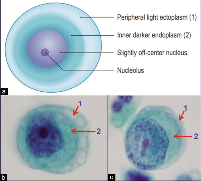
Figure 3:

Mesothelial cells (peritoneal fluid): show outer faintly stained ectoplasm (1) with inner denser endoplasm (2) rich in intermediate filaments. The nucleus is usually central or near central (b), but may be eccentric (c). Nucleoli are readily observed. The vacuolation generally begins at the periphery in ectoplasm (1). [b,c, PAP-stained Autocyte Prep smear (b,c, 100× zoomed).]
