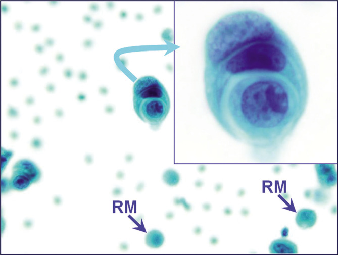
Figure 15:
The cell-in-cell pattern of neoplastic cells (metastatic mammary carcinoma, pleural fluid). A similar pattern may also be observed in reactive mesothelial cells. In this case, the morphology of these cells resembled other cancer cells seen as a second population without overlap with nuclear morphology of reactive mesothelial cells (blue arrows RM). [PAP-stained ThinPrep smear (X100 oil; inset, zoomed)].

