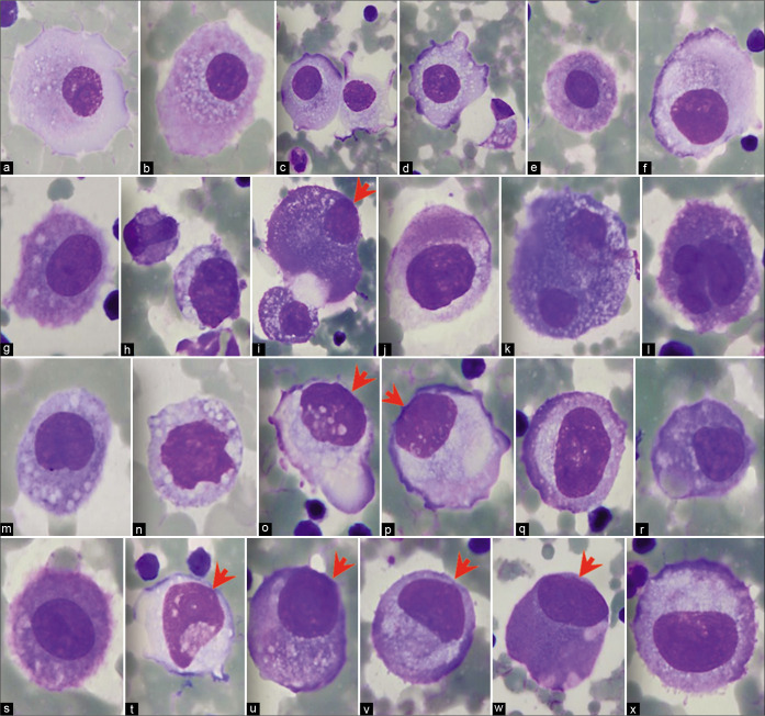
Figure 2:
Panorama of mesothelial cells (ascitic fluid). Central to near central nuclei. Rare mesothelial cells may show eccentric nuclei touching the cell membrane, but usually there is a narrow rim of cytoplasm separating the nucleus from the cell border (arrowheads) [see also Figures 1,3,5]. [a–x, DQ-stained Cytospin smears (a–x, 100X zoomed).]

