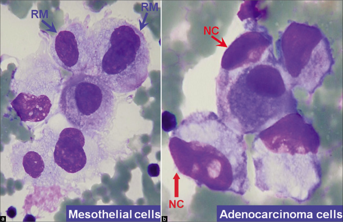
Figure 4:
Mesothelial cells versus adenocarcinoma cells (ascitic fluid). a. Mesothelial cells with central to eccentric nuclei. A thin rim of cytoplasm separates the nuclear border from the cell border (blue arrows RM). b. Compared to mesothelial cells the adenocarcinoma cells with eccentric nuclei appose the cell border without a distinct rim of intervening cytoplasm (red arrows NC). [a,b, DQ-stained Cytospin smear (a,b, 100X zoomed).]

