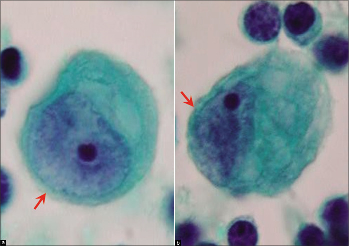
Figure 8:
Mesothelial cells with eccentric nuclei (ascitic fluid); [see more cells in Figure 7b,d,g,h,i,j,m, etc.]. Careful scrutiny usually shows a narrow rim of cytoplasm separating the nucleus from the cell border (arrows). [l,x, PAP-stained ThinPrep smear (100X zoomed).]

