
Figure 2:
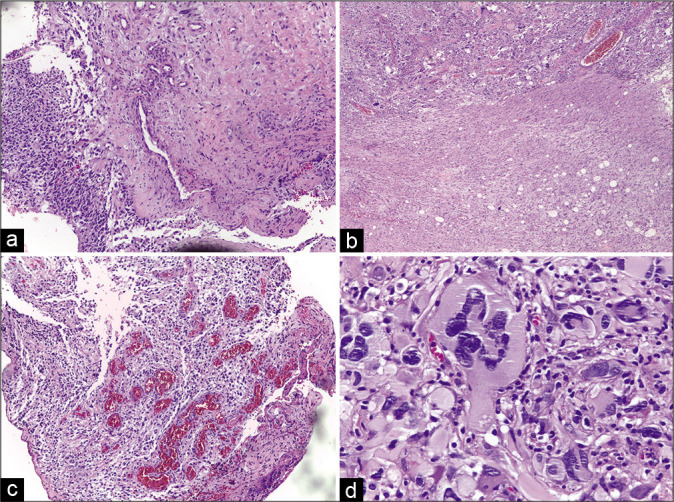
(a-c) Histopathological features of the initial tumor. A routine hematoxylin and eosin-stained specimen shows a pseudopalisading pattern, hypercellularity, and high mitotic proliferation. (d) Nuclear atypia with glomeruloid pattern, typical of glioblastoma.