
Figure 3:
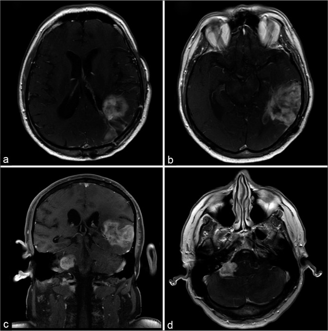
(a and b) Magnetic resonance imaging scans with axial T1-weighted postgadolinium sequence show the initial lesion in the occipital and parietal lobes relapsed. (c and d) The images of the posterior fossa show a new lesion in the contralateral cerebellopontine angle with the same pattern of the supratentorial tumor.