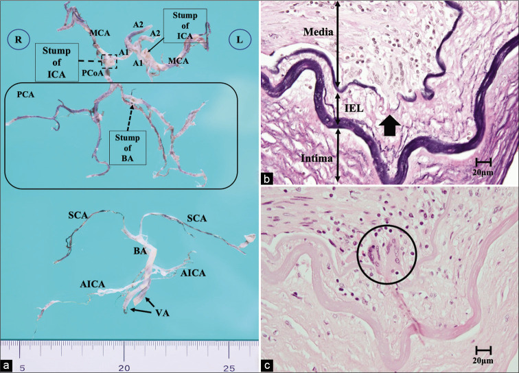
Figure 4:
(a) Macroscopic photograph of the intracranial arteries. The left PCoA was hypoplasia, and the bilateral ICAs were cut at the C2 portion, and the BA was disconnected at the top. (b and c) Microscopic sections of the right ICA at the C2 portion. (b) Elastica van Gieson staining ×400, (c) hematoxylin and eosin staining ×400. All layers of the arterial wall are severely affected by the arteritis, and multinucleated giant cells invade the junction of the media and intima (circle). The IEL was also destroyed (arrow). A1: Anterior cerebral artery A1 segment, A2: Anterior cerebral artery A2 segment, AICA: Anterior inferior cerebellar artery, BA: Basilar artery, IEL: Internal elastic lamina, ICA: Internal carotid artery, MCA: Middle cerebral artery, PCoA: Posterior communicating artery, PCoA: Posterior communicating artery, SCA: Superior cerebellar artery, VA: Vertebral artery.

