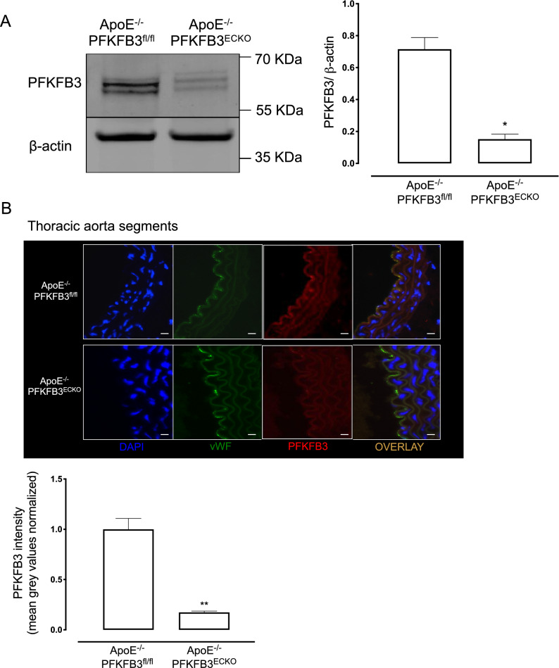
Fig. 2.
Validation of PFKFB3 gene deletion in endothelial cells. A Representative western blot showing protein levels of PFKFB3 and β-actin in primary lung EC. Bars represent relative protein quantification of PFKFB3 normalized to the reference protein β-actin. *P < 0.05 versus ApoE−/−PFKFB3fl/fl (Independent samples t test; n = 3). B Representative thoracic aorta segments of ApoE−/−PFKFB3fl/fl and ApoE−/−PFKFB3ECKO stained with antibodies against PFKFB3 or von Willebrand Factor (vWF; endothelial cell marker). Nuclei were stained with DAPI. Scale bar = 100 μm. PFKFB3 signal in vWF positive cells was quantified. **P < 0.01 versus ApoE−/−PFKFB3fl/fl (Independent samples t test; n = 3)

