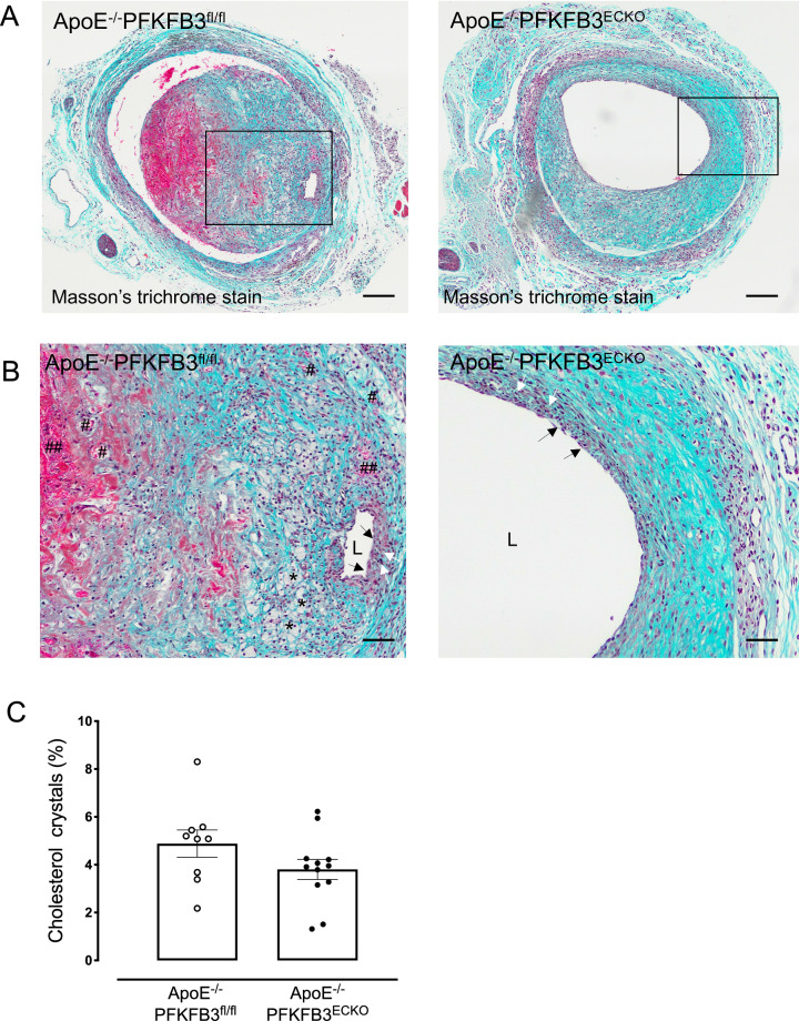
Fig. 3.
PFKFB3 deletion in endothelial cells evokes a more stable vein graft lesion phenotype. A Masson’s Trichrome staining of representative vein grafts. This staining was used for the evaluation of lesion complexity. Nuclei are stained dark blue, erythrocytes are stained bright-red, and collagen is stained blue. The staining allows easy visualization of intraplaque hemorrhages (red areas) as well as necrotic core and cholesterol crystals (white acellular regions). Scale bar = 200 μm. B Boxed area of panel A showing an almost intact endothelium exposed to the lumen (L) (black arrows), a fibrous cap with numerous vascular smooth muscle cells (white arrows), small foam cells (asterisks), an area with extravasated erythrocytes (##), and neovascularization (#). Scale bar = 100 μm. C Quantification of foam cells in vein graft lesions of ApoE−/−PFKFB3fl/fl and ApoE−/−PFKFB3ECKO mice. Independent samples t test; n = 9 (ApoE−/−PFKFB3fl/fl) or n = 12 (ApoE−/−PFKFB3ECKO )

