Fig. 4.
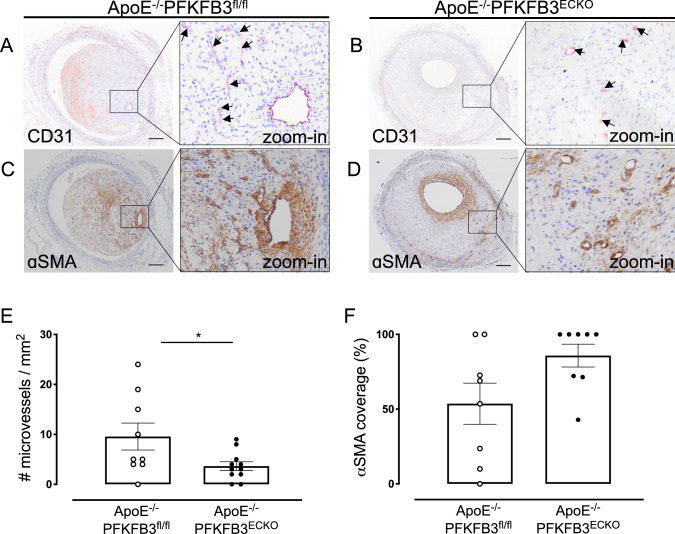
PFKFB3 deficiency in endothelial cells inhibits intraplaque neovascularization in vein graft lesions but does not alter α-SMA coverage of microvessels. A and B Representative vein graft lesions stained with CD31 antibody. Endothelial cells are detected as CD31 positivity (dark violet) around the vessel lumen. Microvessels are marked by arrows. Scale bar = 200 μm. C and D Representative vein graft lesions stained with anti-α-SMA. Vascular smooth muscle cells in the vessel wall are stained by the α-SMA antibodies (brown). E Quantification of microvessels in vein grafts of ApoE−/−PFKFB3fl/fl and ApoE−/−PFKFB3ECKO mice. *P = 0.035 versus ApoE−/−PFKFB3fl/fl. Independent samples t test; n = 9 (ApoE−/−PFKFB3fl/fl) or n = 11 (ApoE−/−PFKFB3ECKO). F Quantification of α-SMA coverage of microvessels in vein grafts of ApoE−/−PFKFB3fl/fl and ApoE−/−PFKFB3ECKO mice. Independent samples t test did not show statistical significance (n = 8 for both groups)

