Fig. 6.
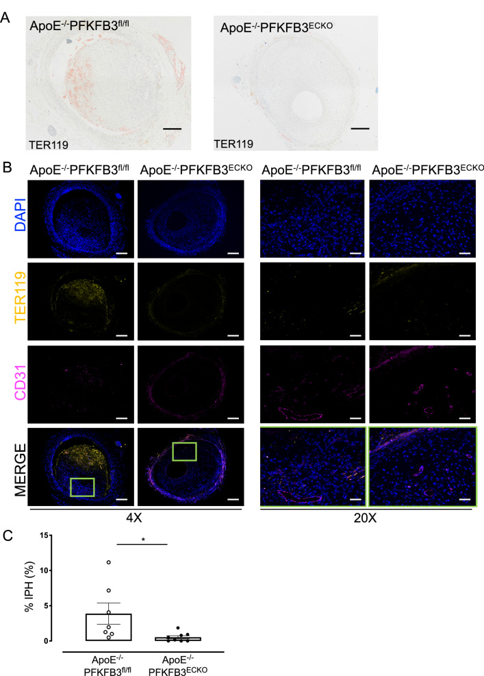
PFKFB3 deficiency in endothelial cells reduces intraplaque hemorrhages (IPH) in vein graft lesions. A Representative vein graft lesions stained with anti-TER-119. TER-119 staining (red) allows detection of erythrocytes. Scale bar = 200 μm. B Examples of atherosclerotic lesions in vein grafts of ApoE−/−PFKFB3fl/fl and ApoE−/−PFKFB3ECKO mice stained with anti-CD31, anti-TER-119, and DAPI. An overlay of the three stainings is also shown as well as a magnification (20x) of the boxed areas showing erythrocyte extravasation. Scale bar = 500 μm (4x) or 100 μm (20x). C Quantification of IPH. *P = 0.034 versus ApoE−/−PFKFB3fl/fl. Independent samples t test; n = 7 (ApoE−/−PFKFB3fl/fl) or n = 8 (ApoE−/−PFKFB3ECKO)

