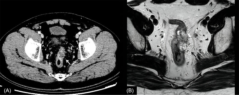
Figure 2.
Rectal cancer with ypT0N0. (A) Axial MSCT depicts a heterogeneous enhancing tumor penetrating the peritoneal reflection (arrow). Over restaged as T4. (B) MRI transverse high-resolution T2WI of rectum shows left mesorectal fascia involvement (arrow). Over restaged as T3N0. yp, pathological staging after neoadjuvant chemoradiotherapy; T, tumor; N, node; MSCT, multi-slice computed tomography; MRI, magnetic resonance imaging; T2WI, T2-weighted imaging.

