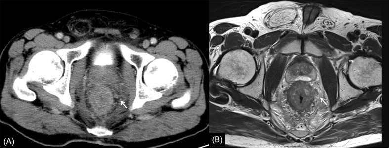
Figure 6.
Rectal cancer with ypT4N2. (A) Axial MSCT depicts whole rectal wall thickening with uneven density of adjacent mesorectum. Low restaged as T3. (B) MRI transverse high-resolution T2WI shows thickened whole rectal wall with extensive edema and fibrosis of mesorectum. Low restaged as T3. Enlarged lymph node (arrow) was found. Accurately restaged as N2 with MSCT and MRI. yp: pathological staging after neoadjuvant chemoradiotherapy; T, tumor; N, node; MSCT, multi-slice computed tomography; MRI, magnetic resonance imaging.

