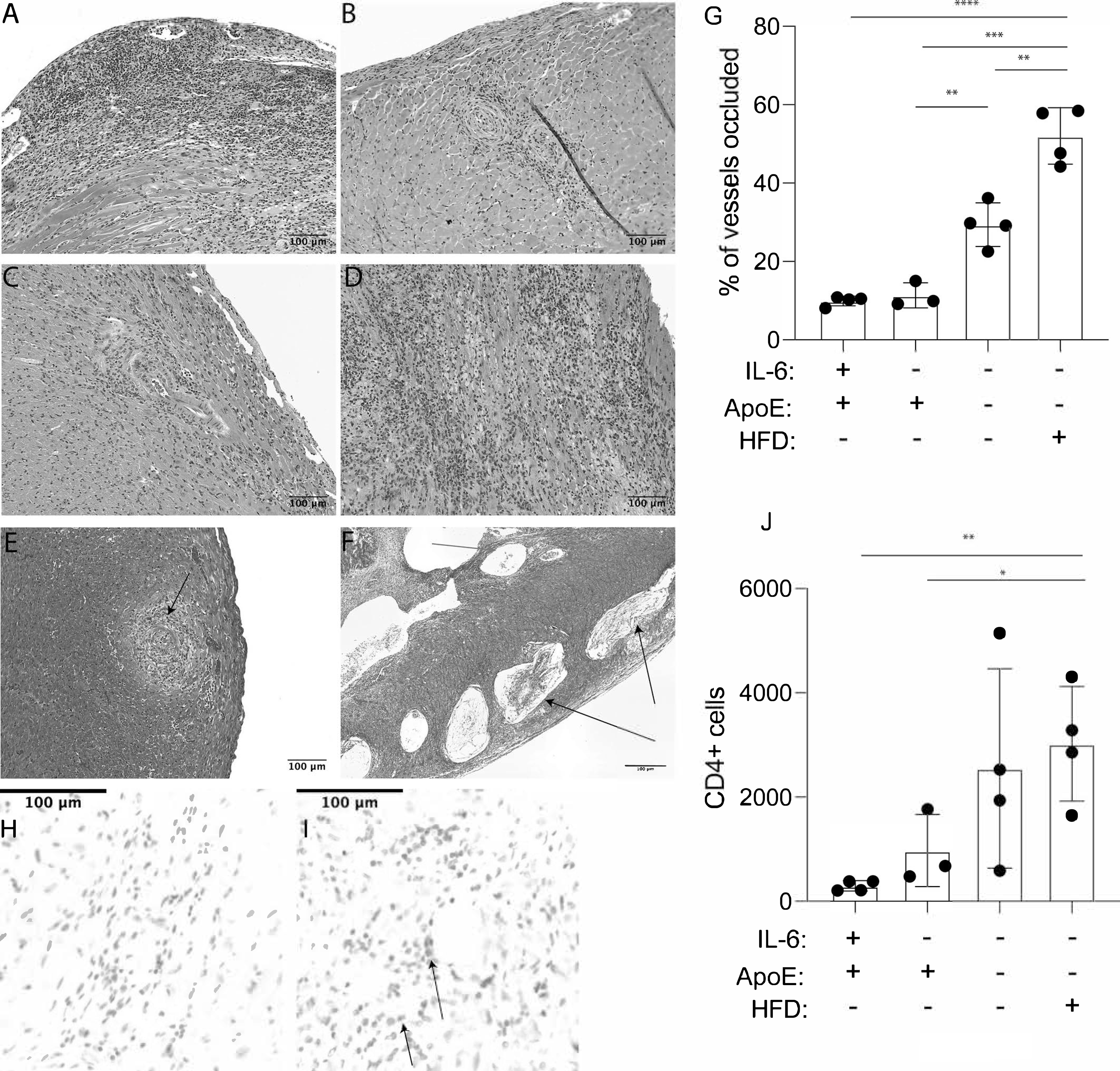
Figure 2. IL-6 independent tissue destruction in cardiac allografts in hyperlipidemic mice.

Age matched groups of mice were fed high fat diet (HFD) or normal chow (NC) for a minimum of four weeks prior to transplant with bm12 cardiac allografts. Grafts were harvested after 100 days, or at the time of rejection, and tissue sections were prepared. Shown are representative sections stained with hematoxylin and eosin (H&E). (A) ApoE−/−IL-6+/+ recipient mice fed HFD (B) ApoE+/+IL-6+/+ recipient mice fed NC (C) ApoE+/+IL-6−/− recipient mice fed NC (D) ApoE−/−IL-6−/− recipient mice fed HFD. Slides were scanned and individual pictures were captured at 15X magnification with CaseViewer software. A representative section showing Masson’s Trichrome staining of occluded vessels and unoccluded vessels in a section from a bm12 graft isolated from (E) ApoE+/+IL-6−/− recipient mice fed NC and (F) ApoE−/−IL-6−/− recipient mice fed HFD. (G) Quantification of occluded vessels in bm12 grafts harvested from recipient animals after 100 days. Tissue sections from 3–4 transplants were analyzed. Statistical significance was determined with a nested t-test. Representative immunohistochemical staining for CD4 in bm12 grafts harvested from (H) ApoE+/+IL-6−/− NC-fed recipient mice (I) ApoE−/−IL-6−/− HFD-fed recipient mice. (J) Quantitation of CD4 cells infiltrating grafts. Tissue sections from 3–4 transplants were analyzed. Error bars represent standard deviation. Statistical analysis was performed using a nested t-Test. **** = P < 0.0001,*** = P < 0.0005, ** = P < 0.005, * = P < 0.05.
