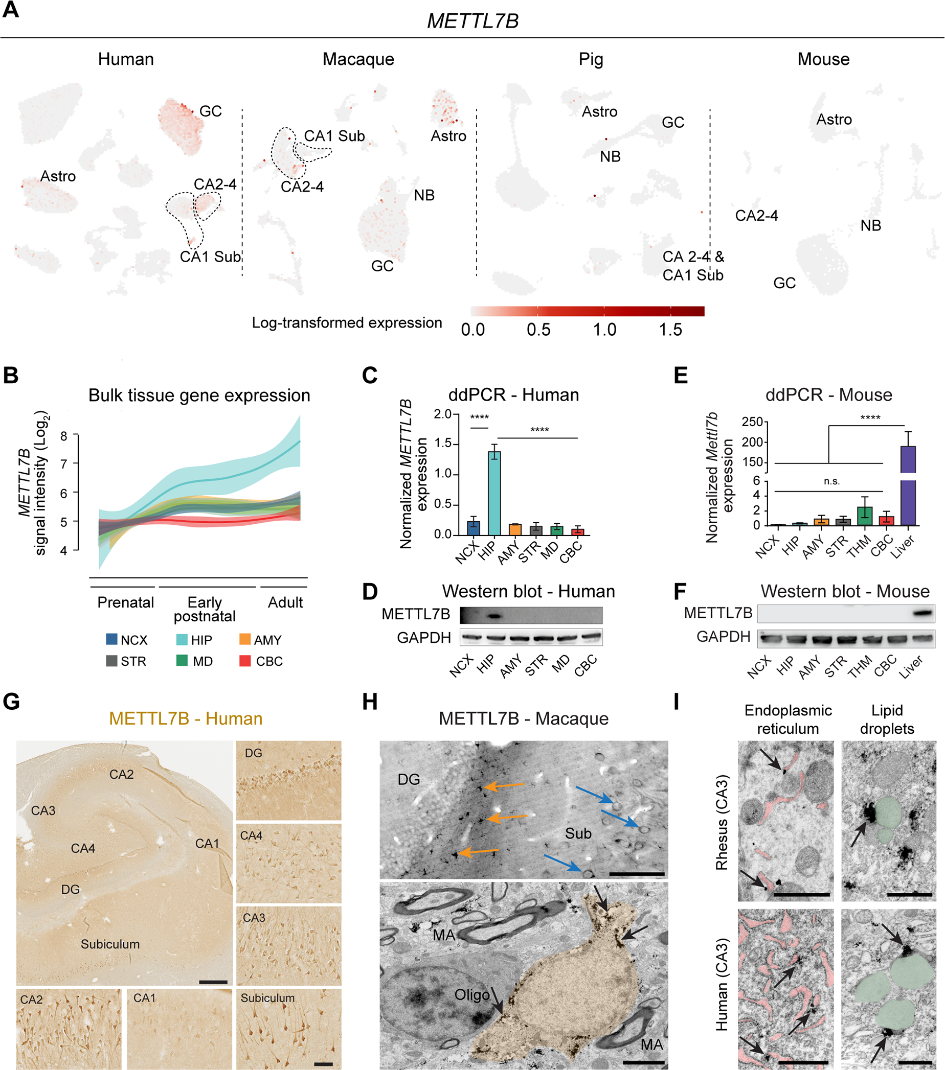
Figure 6. METTL7B defines subregion-specific excitatory neurons and astrocytes in primates.

A, METTL7B expression in adult human HIP-EC, macaque DG, pig HIP and mouse DG. B, Expression of METTL7B showing temporal specificity in adult human hippocampus (Kang et al., 2011). C-D, Droplet digital PCR and immunoblot validation in six regions of adult human brain. One-way ANOVA with post-hoc Dunnett’s adjustment (****P<0.0001), N=3 per group. E-F, Same as (C) and (D) using mouse tissues, including liver as a positive control. G, METTL7B immunostaining of adult human hippocampus. Scale bars = 1 mm; insets = 100 μm; immunofluorescence = 10 μm. H, Upper panel: Numerous METTL7B Immunopositive astrocytes (orange arrows) and neurons (blue arrows). Bottom panel: Immunoelectron microscopy of astrocytes (orange; pointed with arrows). Scale bar is 100 μm (upper) and 2 μm (bottom). MA, myelinated axon. I, Immuno-electron microscopy CA3 hippocampal pyramidal neurons in rhesus macaque and human. Notice METTL7B labeling (arrows) on the outer surface of ER cisterns (pink) and in contact with LDs (green). Scale bar is 1μm for each panel. See also Figure S5.
