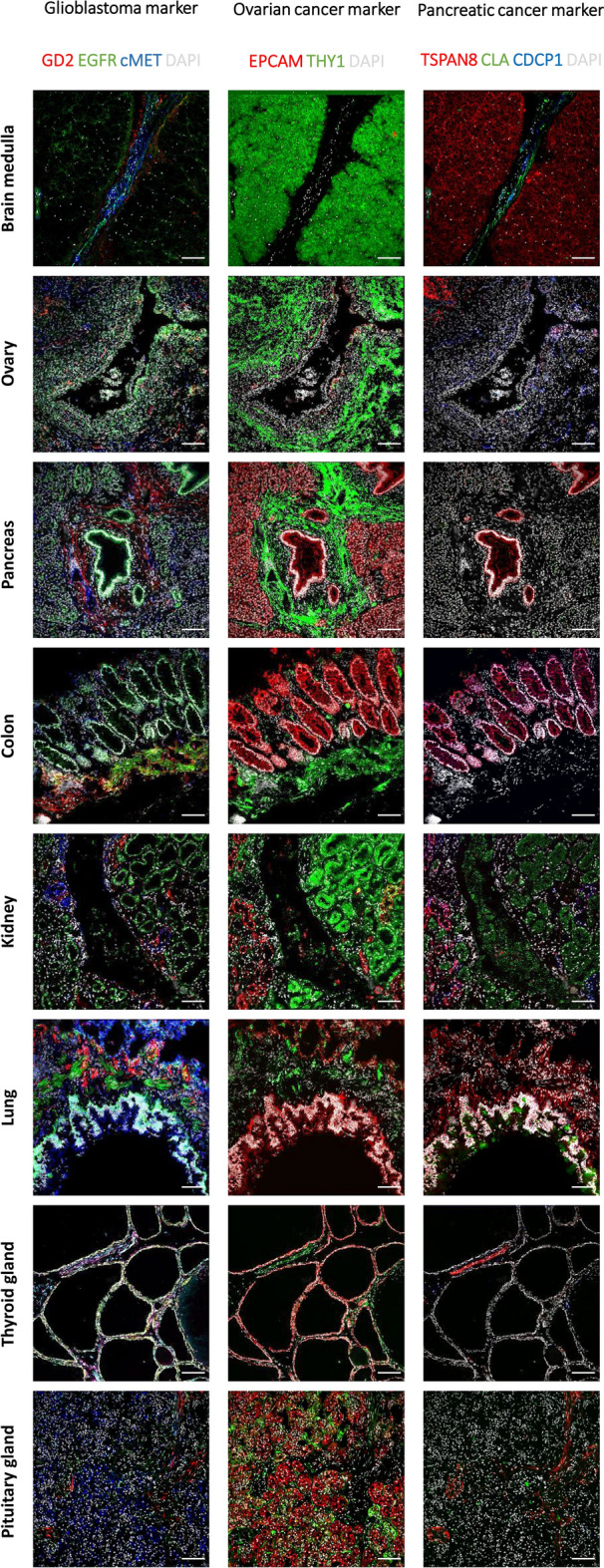
Figure 5.

Ultrahigh-content imaging validates expression of target candidates on healthy human tissues to predict safety and toxicity of target candidates. Fresh-frozen human tissues were sliced and fixed with acetone. The subsequent screening was performed on the MACSima Imaging Platform by employing a sequential staining of antibodies. Healthy human tissues, i.e. medulla oblongata, ovary, pancreas, colon, kidney, lung, thyroid gland, and pituitary gland, were analyzed for the expression of glioblastoma target candidates (left panel), GD2 is shown in red, EGFR in green, cMET in blue, and DAPI in white; ovarian cancer target candidates (middle panel), EPCAM is shown in red, THY1 in green, and DAPI in white; pancreatic cancer target candidates (right panel), TSPAN8 is shown in red, CLA in green, CDCP1 in blue, and DAPI in white. Scale bar represents 100 µm.
