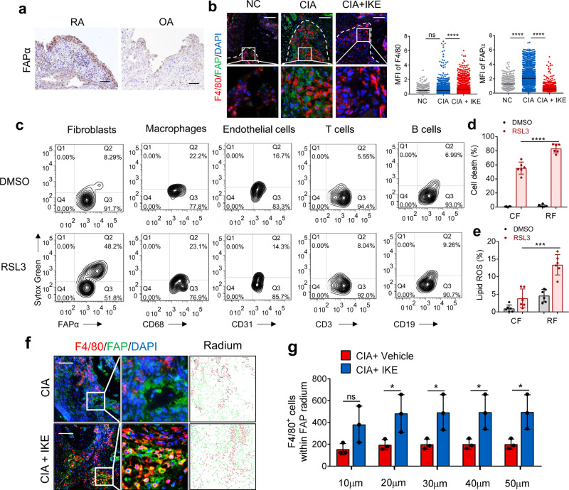
Fig. 2. The ferroptosis inducer IKE decreased fibroblast populations in RA synovium.
a Representative immunohistochemical staining of FAPα in hyperplastic rheumatoid synovium of 26 RA and 21 OA patients. Scale bars, 50 μm. b Representative fluorescent multiplex IHC staining and scoring of joints labeled with anti-F4/80 (red), anti-FAPα (green), and DAPI (blue). Scale bars, 100 μm. n = 413 (NC), 3698 (CIA), 3469 (CIA + IKE) cells from 3 independent joints for each group. ****P < 0.0001; ns, P = 0.6849; one-way ANOVA followed by multiple comparisons. c Cell death in FAPα+ fibroblasts, CD68+ macrophages, CD31+ endothelial cells, CD3+ T cells, or CD19+ B cells isolated from hyperplastic synovium treated with 0.125 μM RSL3 for 6 h, quantified by SYTOX staining followed by flow cytometry. Cell death (d) and lipid ROS production (e) in circulating fibrocytes from PBMCs and synovial fibroblasts from inflamed joint fluid of RA patients treated with 0.125 μM RSL3 for 18 h (cell death) or 6 h (lipid ROS). ****P < 0.0001, ***P = 0.0001; two-tailed t-test. n = 6 patients. f Representative fluorescent multiplex IHC staining of retained hyperplasia synovium of CIA model mice treated with or without IKE and labeled with anti-F4/80 antibody (red), anti-FAPα antibody (green), and DAPI (blue). Scale bars, 200 μm. g The number of F4/80+ macrophages within the 50 μm radium of FAPα+ fibroblasts in joints of CIA mice. *P < 0.05; ns, P = 0.00878; two-tailed t-test. n = 3 joints. Data are presented as mean ± SD. Source data are provided as a Source data file.

