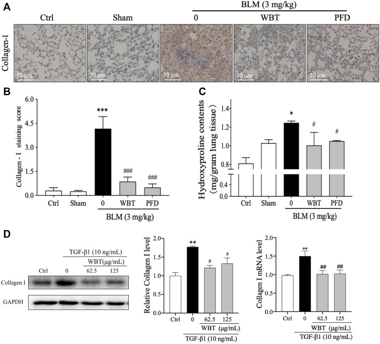
FIGURE 7.
WBT formula inhibited BLM-induced collagen deposition in the mouse and TC-1 cell model of lung fibrosis. (A) The level of collagen I was detected by immunohistochemical staining in the mouse lung from different groups. Scale bar = 50 μm. (B) The relative collagen I level from (A) was quantified (n = 5); ***p < 0.001, compared to the sham group; ###p < 0.001, compared to the BLM group. (C) The content of hydroxyproline in the lung tissues from different groups was determined by a commercial measurement kit. (D) After incubation with TGF-β1 and/or WBT for 48 h, qPCR and Western blot analysis were performed. The relative levels of collagen I in TC-1 cells were calculated and normalized to GAPDH. Data are from three independent experiments. *p < 0.05, **p < 0.01, and ***p < 0.001, compared to the sham group; #p < 0.05, ##p < 0.05, and ###p < 0.001, compared to the BLM or TGF-β1 group. BLM, bleomycin; WBT, Wenfei Buqi Tongluo formula; PFD, pirfenidone (TGF-β1, 10 ng/ml).

