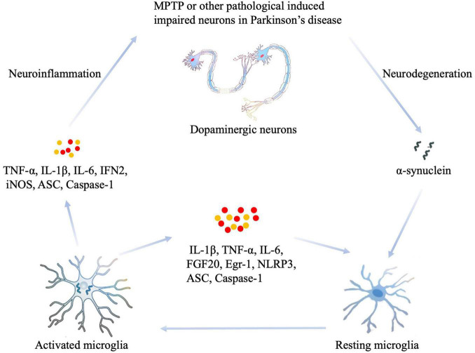
FIGURE 1.
The role of MG in the neuroinflammation that occurs during PD. Damaged or dying neurons in PD, or toxin-induced models such as MPTP, cause the release of impair-associated molecular patterns (IAMPs) and α-synuclein to activate microglia through pattern recognition receptors (PRRs). Thus, the resting states of MG can be activated, thus leading to the eventual release of proinflammatory cytokines, including IL-1β, IL-6, TNF- α, and other inflammatory mediators such as ASC, IFN2, iNOS, and Caspase-1. In addition, increased levels of proinflammatory cytokines activate other resting MG, which induces neuroinflammation or neurodegeneration in the damaged area. MPTP, 1-methyl-4-phenyl-1,2,3,6-tetrahydropyridine; IL-1β, interleukin-1β; IL-6, interleukin-6; IFN2, type 2 interferon; iNOS, inducible NOS; TNF-α, tumor necrosis factor-α; ASC, apoptosis-associated speck-like protein containing a CARD.

