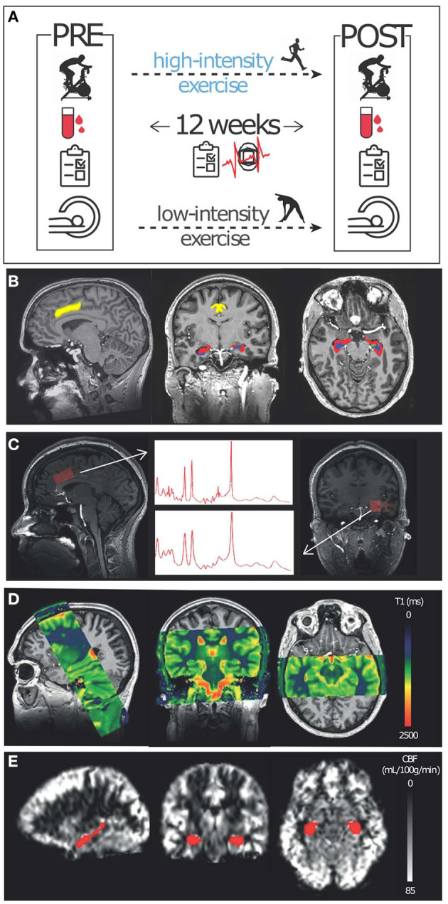
Figure 2.

Study methods: (A) Participants were enrolled in a 12-week low- (active control) and high-intensity exercise intervention. Several measures, including a cardiorespiratory fitness test (VO2max), and peripheral growth factors (blood sampling) were conducted before (PRE) and after (POST) the exercise intervention. Additionally, HR, exercise frequency, and exercise questionnaires were collected during the intervention. Furthermore, several MRI measures were collected before and after the exercise regime: (B) T1- and T2-weighted scans were conducted at 7T for segmentation purposes. (C) Single voxel spectroscopy was conducted at 7T in the dACC (left) and left hippocampus (right). (D) T1-mapping using a steady-state contrast-enhanced method was conducted at 3T to derive CBV and R1. (E) A pCASL sequence was used at 3T to obtain CBF values.
