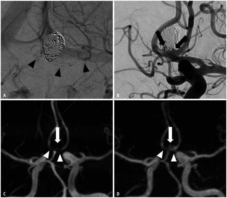
Fig. 3. Images of a 42-year-old male with a treated aneurysm in the anterior communicating artery.
A. Stent-assisted coil embolization was performed using an LVIS Jr stent. Arrowheads indicate implanted stent markers. B. Final angiography during the embolization procedure shows the neck remnant (arrows) of the aneurysm sac. C. Time-of-flight MRA shows acceptable visualization at the stented segment of the anterior cerebral artery (arrowheads), and the neck remnant is not depicted (arrow). D. Silent MRA shows excellent visualization at the stented segment (arrowheads), and the neck remnant is fully depicted (arrow). MRA = MR angiography

