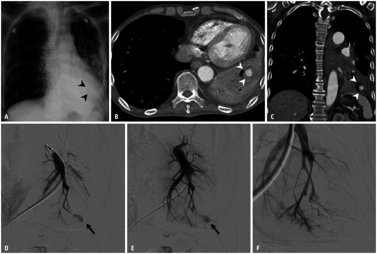
Fig. 11. Pulmonary artery pseudoaneurysm in a 65-year-old male with lung cancer undergoing chemotherapy.
A. Chest radiograph shows a thin, radiolucent crescent-shaped structure (arrowheads) superimposed onto the enlarged cardiac silhouette. B, C. Axial and coronal CT images reveal an air crescent adjacent to a pseudoaneurysm (arrowheads) with surrounding consolidation. D, E. Selective angiogram of the left lower lobe pulmonary artery demonstrates the pseudoaneurysm (arrows) arising from the segmental branch. F. Completion angiogram after coil embolization shows the absence of pseudoaneurysm filling.

