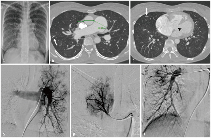
Fig. 12. Pulmonary artery ectasia in a 56-year-old female patient with hemoptysis.
A. Chest radiographs show mild cardiomegaly and prominent bilateral pulmonary vascular markings. B, C. Axial images in the lung window setting show dilatation of the pulmonary trunk to a maximum diameter of 36 mm with dilated and beaded peripheral pulmonary arteries (arrows). A ventricular septal defect is also noted (arrowhead). D-F. Arterial-phase images of the selective bilateral pulmonary angiogram reveal diffuse dilatation of the pulmonary trunk and branches.

