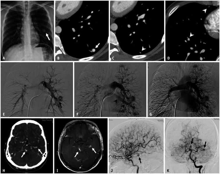
Fig. 2. A 19-year-old male patient with hereditary hemorrhagic telangiectasia.
A. Chest radiograph shows a well-circumscribed round opacity (arrow) in the lower third of the left hemithorax. B-D. Axial enhanced chest CT images demonstrate large arteriovenous malformation (arrow) with a feeding artery in the left lower lobe. Numerous, small arteriovenous malformations (arrowheads) are also identified in both lung fields. E, F. Arterial-phase images from the pulmonary angiogram reveal the feeding artery (arrow) and rapid opacification of the draining vein (arrowheads). G. Delayed venous-phase image shows opacification of the normal pulmonary veins. H, I. Contrast-enhanced brain CT and contrast-enhanced T1-weighted MR image demonstrate two small enhancing lesions (arrows) located at the left lateral ventricle temporal horn and midbrain, which suggest brain arteriovenous malformation. J, K. Left cerebral and vertebral angiograms confirm two small arteriovenous malformations (arrows).

