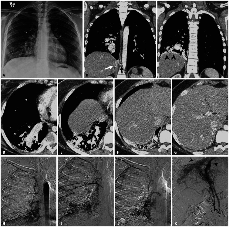
Fig. 4. A 19-year-old female patient with pleural arteriovenous malformation who underwent pulmonary artery embolization six years earlier.
A. Chest radiograph shows markedly enlarged vascular markings at the right lower lung zone. Multiple plugs from prior embolization procedures were also noted. B-G. Axial and coronal contrast-enhanced CT images with a mediastinal window setting demonstrate engorged vascular structures along the diaphragmatic pleural surface (arrowheads) and extremely dilated right inferior pulmonary veins. The feeding arteries originated from the hypertrophied right intercostal (black arrow) and inferior phrenic arteries (white arrow). H-K. Thoracic aortogram and selective angiograms of the right intercostal and inferior phrenic arteries reveal pleural arteriovenous malformation and early drainage through dilated right inferior pulmonary veins (arrowheads).

