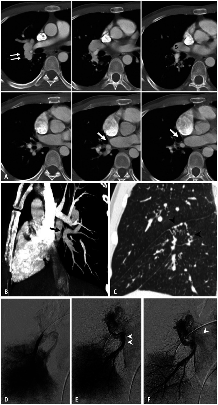
Fig. 5. An anomalous pulmonary vein caused by lobar pulmonary vein atresia in a 32-year-old male patient.
Adapted from Lee et al. Korean J Radiol 2011;12:395-399 [30].
A. Serial axial CT images show a low attenuation linear structure (arrows) between the right inferior pulmonary vein and left atrium, as well as a prominent right superior pulmonary vein(s). An abnormally enlarged vascular structure (double arrow) near the right pulmonary artery, suggesting the presence of collateral drainage of the interrupted inferior pulmonary vein, was also noted. B. The reformatted oblique sagittal CT image shows the atretic right inferior pulmonary vein (arrow). C. The reformatted coronal image with the lung setting reveals multiple tortuous and dot-like collaterals (arrowheads) in the superior segment of the right lower lobe crossing the major fissure. D-F. Venous-phase images of the right interlobar selective pulmonary angiogram show no connection between the left atrium and the right inferior pulmonary vein (double arrowhead), which is draining into the superior pulmonary vein (arrowhead) through the collaterals.

