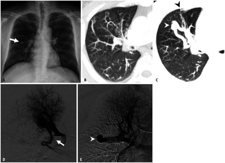
Fig. 7. An anomalous pulmonary vein and segmental pulmonary vein atresia in a 43-year-old male patient who presented with persistent cough and sputum production.
Adapted from Lee et al. Korean J Radiol 2011;12:395-399 [30].
A. Chest radiograph shows a single nodular opacity (arrow) in the right mid-lung zone. B, C. Slab axial images with the lung window setting show tortuous and dot-like collaterals and an aneurysmal dilatation of vascular structure (white arrowhead) crossing the minor fissure (black arrowhead) in the anterior segment of the right upper lobe. D, E. Venous-phase images in the right apical segmental selective pulmonary angiogram show that the apical segmental pulmonary vein is interrupted (arrow). Venous drainage of the affected segment detours via pulmonary vein varix associated with anomalous pulmonary vein (arrowhead).

