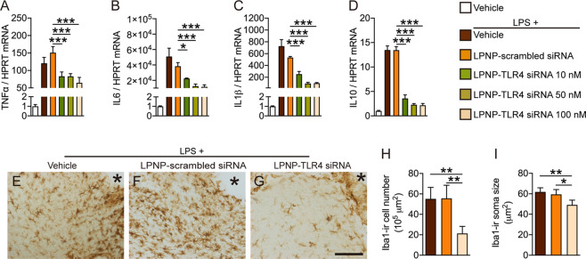
Figure 6.
LPNP-TLR4 siRNA downregulates the immune response of microglia following LPS stimulation. (A–D) Gene expression of TNF-α, IL-6, IL-1β, and IL-10 in cultured microglial cells treated for 24 h with LPNP-TLR4 siRNA (n = 3) at different concentrations (0, 10, 50, and 100 nM, with 0 nM representing LPNP-scrambled siRNA (100 nM)). Afterward, the cells were treated with LPS (100 ng/mL) for 2 h (in addition to the first PBS group). (E–G) Representative images show Iba1-ir microglial cells near the injection spot (asterisk) at the morphological level in the rat hypothalamus treated with LPNP-TLR4 siRNA 18 h before followed by intravenous LPS (100 μg/kg) 2 h before sacrifice. (H) Quantification of the Iba1-ir microglial cell number and (I) soma size following LPS stimulation (n = 4). The data are presented as means ± SD and were analyzed using one-way ANOVA, *p < 0.05; **p < 0.01; ***p < 0.001. Scale bar: 100 μm in panels (E–G).

