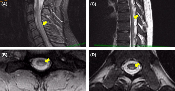
FIGURE 2.

T2‐weighted magnetic resonance (MR) imaging. Sagittal slice of the cervical spine with diffuse hyperintensity of the dorsal column and mid cord (A). Axial slice of the cervical spine with diffuse hyperintensity of dorsal column and mid cord (B). Sagittal slice of the thoracic spine with diffuse hyperintensity of the dorsal column (C). Axial slice of the thoracic spine with symmetric hyperintensity of the dorsal column (D)
